Soft Agar Assay
In temperature responsive gel

3D Gel for Adhesion Free Cell Culture
poly(2-3)Glucose™ T1-35 temperature responsive gel replaces soft agar, and methylcellulose media in anchorage independence and colony formation assays. Cells do not adhere to the biocompatible gel, so 3D cell culture selects for metastatic tumor cells or for differentiation of chondrocytes and stem cells. The advantage of 3D cultures over 2D low adhesion substrates is colony growth from a single cell dispersed in the gel instead of growth of a spheroid from a clump of cells that were aggregated on the substrate. The advantages of poly(2-3)Glucose™ for tumorigenicity and clonogenic assays over soft agar and methylcellulose is the easy, gentle recovery of cells for subsequent omics analysis, cell counting, or replating. T1-35 is a liquid below 35 C and a 3D cell culture gel at 37 C, so cells are handled in suspension at room temperature, heated only to 37 C for culture, and then chilled for recovery. Melting the gel by chilling enables seeding/establishing the cells for 24-48 hours at 37 C, then dissolving the gel to thoroughly mix in chemotherapeutics, and returning to gel culture at 37 C. Anchorage-independent growth, a marker of malignant transformation, and clonogenic assays for cell differentiation can grow on ultra-low adhesion surfaces or in 3D artificial matrices like soft agar and methylcellulose, but temperature responsive gel provides the least cell manipulation and the gentlest recovery of transformed cells.
3D matrices represent physiologically relevant models of cellular behavior, as opposed to 2D growth, but cell recovery is a significant problem for soft agar. The 42 C temperature required for gelling agar during assay assembly introduces experimental variables that can make interpretation of the results problematic. In the effort to add the cells to the agar at the lowest temperature possible, premature hardening of the agar can lead to incomplete and inhomogenous seeding. The 80 C melting temperature of agar makes cell recovery impossible. Methylcellulose media do not gel, but semi-solid media force a trade-off between creating enough elasticity to mimic the extracellular matrix, and too much shear stress while handling the cells. poly(2-3)Glucose™ allows cells to be gently handled in a low viscosity liquid, grown in a soft gel, and recovered by brief chilling.
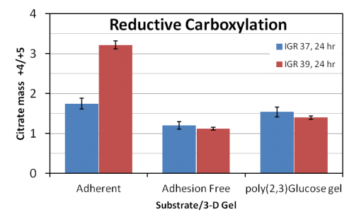
Metabolic pool sizes and post translational modifications are sensitive to manipulation during harvesting, so recovery with chilling enables metabolomic and proteomic analysis of transformed cells. poly(2-3)Glucose™ T1-35 liquefies by brief chilling on ice, conditions in which all biological activity is significantly slowed. Cells are recovered in an ideal way that preserves intracellular metabolite composition and amounts as well as “freezing” the patterns of PTMs, gene expression. The figure (below, top) illustrate metabolomic analysis of 13C glucose conversion to citrate. The data show similar metabolic results from adhesion free substrates and poly(2-3)Glucose™ T1-35.
Soft agar displays temperature hysteresis. It gels at 42 C, but it melts at 80 C so cells cannot be recovered. Cells must be added as close to 42C as possible, so the gel sets very rapidly and wells can be difficult to fill evenly. poly (2-3)Glucose™ T1-35 is liquid at temperatures below 35 C. So it is easy to fill wells at room temperature and to recover cells by chilling. Temperature responsive gel is faster to assemble than agar because the cells in polymer solution are handled at room temperature. No special concern is needed for handling agar near its solidification point.
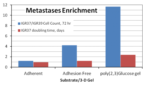
Once in culture, T1-35 enriches metastatic over primary tumor cells. The figure (below, bottom) shows 12x increase of metastatic (IGR37) melanoma cell count over primary (IGR39) tumor cell count in 72 hours. Slower growing cells can also be recovered in 7 days and measured by MTT assay instead of waiting for colony formation. The recovered spheroids can also be replated.
- Add sterile medium through septa on T1-35 vial
- Dissolve T1-35 o/n in refrigerator
- Mix and add cells at RT
- Fill well plate, gels in seconds at 37 C
- Handle the gel at RT for hours
- Recover cells in 10-15 min in ice bath
poly(2-3)Glucose™ 3D Cell Culture Protocol
The procedure described herein is for a 6-well plate format (Costar cat # 3516, Corning Corporation).
- Reconstitute the T1-35 gel at 5% w/v in MEM, 10% FBS and vortex briefly to allow the gel cake to hydrate. One well of the plate requires 2 mL total of the gel solution.
- Place the reconstituted gel at 2-8ºC overnight.
- The next day pour the liquefied gel at 1 mL per well and place at 37º C for 2-3 hours.
- After the gel in the plate solidifies (turns opaque), prepare cell suspension in the remaining gel solution and immediately pour on top of the solidified gel surface.
- Place the plate in the 37ºC incubator for the duration of the experiment. After the layer containing cells solidifies, cells can be viewed under the microscope. If needed growth medium can be added on top of the gel.
- To extract live cell colonies for analysis, place the plate on ice for 40-45 minutes. Make sure the gel is completely liquefied before collecting the cells. Cells can be pelleted by centrifugation and the liquid gel material can be either collected for analysis or discarded.
Table 1 Recommended use of poly(2-3)Glucose polymer T1-35 for the assay
| Corning | Bottom layer | Top layer | Total vol | Total vol | Total T1-35 |
| Well per plate | mL per well | mL per well | mL per well | mL per plate | gm per plate |
| 6 | 0.8 | 0.8 | 1.6 | 9.6 | 0.48 |
| 12 | 0.4 | 0.4 | 0.8 | 9.6 | 0.48 |
| 24 | 0.2 | 0.2 | 0.4 | 9.6 | 0.48 |
| 48 | 0.1 | 0.1 | 0.2 | 9.6 | 0.48 |
| 96 | 0.05 | 0.05 | 0.1 | 9.6 | 0.48 |
Rapid cell recovery at 0<sup>o</sup> C
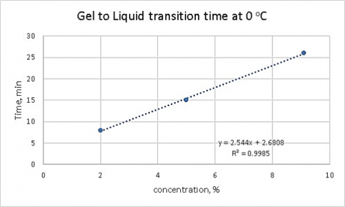
Gel temperature

Handle the gel at RT